2019B15 Describe the anatomy and relations of the right
internal jugular vein relevant to performing
central venous cannulation.
List:
· Diagramme
· Anatomy description: location, passage, borders
· Approach: landmark and ultrasound
· Complications: especially those relevant to anatomy
Diagramme:
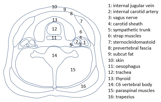
Anatomy:
|
Location |
In the anterior triangle · Anterior midline · Anterior border of sternocleidomastoid · Inferior angle of the mandible |
|
Origin |
· Confluence of R sigmoid sinus + R inferior petrosal sinus, at the jugular foramen |
|
Passage |
· Passes vertically down the neck · Within the carotid sheath |
|
Termination |
· R internal jugular vein + R subclavian vein -> R brachiocephalic vein · Note direct continuation to SVC and right atrium, hence right internal jugular preferred to left |
|
Relation to internal carotid artery |
· At C2: posterior · At C3: posterolateral · From C4: lateral · (note may appear posterior in the lower neck if ultrasound applied radially rather than antero-posterior) |
|
Relation to other major neck structures |
· Anterior: sternocleidomastoid · Posterior: lateral mass of C1, anterior and middle scalenes, pleura of lung apices · Medial: thyroid, trachea, oesophagus · Lateral: sternocleidomastoid, fascia, skin |
Approach:
|
Landmark |
· Trendelenburg (prevent air embolus) · Head rotated contralaterally · Palpate carotid artery medially · Enter skin in the middle of the triangle formed by the two heads of SCM and clavicle (~C6 level) · Angle needle 30 degrees to the skin · Aim at ipsilateral nipple |
|
Ultrasound |
· Gold standard · Same prep + position · Probe best positioned for AP view rather than radial view to avoid carotid puncture · Identify vessels by shape, pulsatility, compressibility, direction of blood flow |
Complications:
|
Damage to surrounds |
· Common carotid artery or internal carotid artery -> bleeding, stroke · Vagus nerve · SNS nerve -> Horner’s syndrome · Pleura -> pneumothorax · Trachea, oesophagus, thyroid · Thoracic duct -> chylothorax |
|
Other |
· Arrythmia (wire in RV) -> myocardial ischaemia if susceptible · Venous air embolism · Bleeding · Infection |