2020B06 Draw and label a normal capnograph to show the phases of the
respiratory cycle (30%).
List and briefly describe what information can be obtained from the capnograph
(70%).
List:
· Diagramme
· Components
· Airway & breathing
· Circulation
· Other
Diagramme:
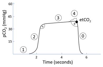
Components:
|
1 |
Baseline |
· Should be near 0mmHg |
|
2 |
Expiratory upstroke |
· Transition from anatomical dead space to alveolar gas |
|
3 |
Alveolar plateau |
· Alveolar gas |
|
4 |
Terminal upswing |
· Rarely present |
|
* |
etCO2 |
· Approximates PaCO2 |
|
0 |
Inspiratory downstroke |
· Early inspiration |
|
α |
Angle |
· Transition between phases 2 and 3 |
|
β |
Angle |
· Transition between phases 3 and 0 |
Airway & Breathing:
|
ETT placement |
· Trachea: normal waveform · Bronchus: ± bifid waveform (delayed expiration from other lung) · Oesophagus: small, vanishing sinusoidal waveform · Disconnection: no waveform |
|
Ventilation |
· Mode of ventilation o Spontaneous: ↑slope phases 0 and 2, shortened phase 3 o Mechanical: square-ish waveform, longer phase 3 · Adequacy of ventilation: o VA ∝ 1/ etCO2 · Alveolar dead space o ∝ difference between etCO2 to PaCO2 (from ABG) o Normal 2-5mmHg · Rebreathing: ↑phases 1 and 3 o Important for Mapleson classification circuits · Bronchospasm: ↑slope phase 3 (and loss of α angle) o Increased heterogeneity of time constants o Earlier emptying of fast lung unit with low pCO2 o Later emptying of slow lung units with high pCO2 o Worse if short expiratory time · Emphysema: reversal of slope phase 3 (and ↑β angle) o ↑↑Alveolar volume, early completion of gas exchange · Small airway closure: terminal upswing = pigtail o Closure of lung units with low pCO2 o Continued emptying of lung units with high pCO2 o Seen in pregnancy, obesity, poor compliance (e.g. ARDS) |
|
Equipment dysfunction |
Tubing: · Circuit obstructed externally: ↓slope phases 0 and 2 · Circuit disconnected: no waveform Valves: · Insp valve stuck open: ↓slope phase 1 · Insp valve stuck closed: no waveform · Exp valve stuck open: ↑phases 1 and 3 · Exp valve stuck closed: ↓etCO2 (once ventilation fails) Sampling line: · Leak -> ↓early phase 3, ↑terminal phase 3 (= dual plateau) · ↑Pressure at onset of inspiration reduces air entrainment |
Circulation:
|
Cardiac output (RV) |
· Low output: ↓phase 3 · Arrest without CPR: no waveform · Arrest with CPR: etCO2 >20mmHg indicates effective CPR |
|
Cardiac oscillations |
· Small, regular, rapid sinusoidal waveforms · Seen in low frequency ventilation · Cardiac impulse causes back and forth motion between exhaled and fresh gas |
Other:
|
Muscle relaxation |
· Spontaneous inspiration produces ‘curare cleft’ in phase 3 |
|
Sodalime exhaustion |
· ↑Phase 1 and 3 for a given MV · Normalises if ↑FGF |
|
Metabolic rate |
· MR ∝ VCO2 ∝ height of Phase 3 o ↑MR: sepsis, MH o ↓MR: hypothermia |
Feedback welcome at ketaminenightmares@gmail.com