2006B13 Briefly
describe the structure of a mammalian skeletal muscle fibre and explain how
its structure is related to its contractile function. DO NOT describe
excitation-contraction coupling.
List:
· Definitions
· Membrane structures
· Contractile structures
· Other intracellular structures
Definitions:
· Muscle = entire structure
· Myocyte = muscle fibre = single cell, 10-100 micrometres diameter
· Myofibril = bunch of contractile elements, many in each myocyte
· Myofilament = individual contractile protein
Membrane structures:
|
Neuromuscular junction |
· Link between somatic nervous system and skeletal muscle · Single interface allows co-ordinated activity · Junctional nAChR: for rapid depolarisation (millions of ACh molecules and receptors) · Peri- and extra-junctional VDNaC: to start and propagate action potential |
|
Sarcolemma |
· Lipid bilayer · Invaginations (T tubules) extend towards sarcoplasmic reticulum, ensure synchronous contraction · Abundant ion channels, especially VDNaC, VDKC, Na+K+ATPase, ensure synchronous contraction |
Contractile structures:
|
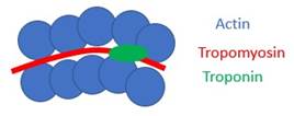
Components |
· Actin = thin filament · Myosin = thick filament, has contractile heads · Tropomyosin = fits in groove between actin monomers, prevents interaction with actin · Troponin:
controls tropomyosin, responds to change in ICF [Ca2+]
|
|
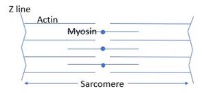
Sarcomere |
· Parallel relationship means additive force production in a single direction |
Other intracellular structures:
|
Nuclei |
· Multiple due to fusion of myoblasts |
|
Mitochondria |
· For aerobic metabolism · More abundant in slow twitch oxidative |
|
Sarcoplasmic reticulum |
· Analogous to the smooth endoplasmic reticulum · Calcium reservoir · Ion channels = ryanodine channel · Allows rapid change in ICF [Ca2+] to start and end contraction |
|
Glycolytic enzymes |
· For anaerobic metabolism and start of aerobic ATP production |
|
Myoglobin |
· Binds oxygen with very high affinity (p50 2.8mmHg) · Preserves concentration gradient in peak exercise · More abundant in slow twitch fibres |